
ApaI Restriction Enzyme: Recognition Site, DNA Digestion and Cloning Applications
ApaI Restriction Enzyme: DNA Recognition, Rapid Digestion and Molecular Cloning
A detailed scientific guide to ApaI restriction endonuclease activity, GG GCCC recognition, neoschizomers Bsp120I and PspOMI, methylation sensitivity, DNA cloning, and the JBSpeed digestion workflow.
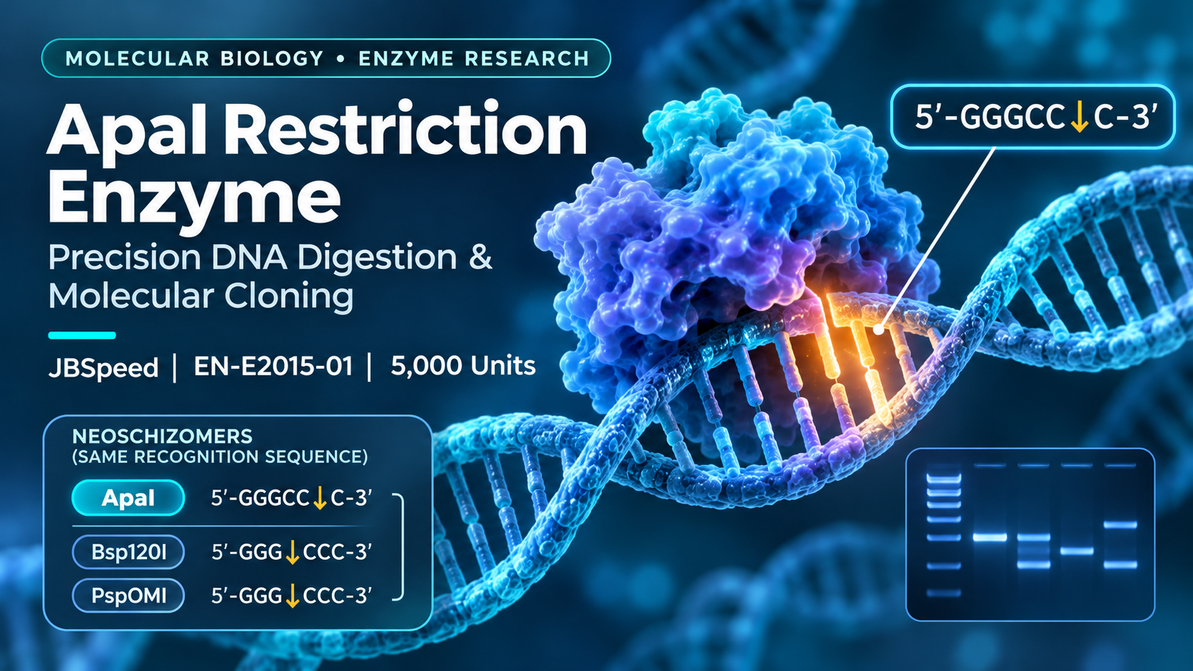
ApaI is a sequence-specific type II restriction endonuclease that recognizes the palindromic double-stranded DNA sequence 5′-GGGCCC-3′. It cleaves within this recognition site to generate four-nucleotide 3′ cohesive ends.
ApaI has two important neoschizomers, Bsp120I and PspOMI. These enzymes recognize the same sequence but cut at different positions, producing different DNA termini.
The Jena Bioscience JBSpeed ApaI formulation is designed for rapid DNA digestion using the recommended Universal Buffer and a short incubation at 25°C.
What Are Restriction Endonucleases and How Do They Work?
Restriction endonucleases are enzymes that recognize particular DNA sequences and catalyze the cleavage of phosphodiester bonds within or near those sites.
They are fundamental tools in molecular biology because they enable controlled fragmentation of double-stranded DNA, supporting recombinant DNA construction, restriction mapping and genetic analysis.
Type II Restriction Enzymes
Many commonly used laboratory restriction enzymes belong to the type II family. These enzymes typically recognize short, defined DNA sequences and cleave DNA at reproducible positions.
Recognition sites are often palindromic: their sequences read identically in the 5′-to-3′ direction on both complementary strands.
What Makes ApaI Important?
ApaI recognizes a specific six-base DNA sequence and produces cohesive DNA termini that can be useful in molecular cloning applications.
ApaI recognizes a defined six-base DNA sequence, GGGCCC.
Cleavage produces four-base 3′ single-stranded extensions.
Sequence-specific DNA cleavage supports recombinant DNA workflows.
Restriction Enzymes in Modern Research
Restriction enzymes remain useful despite the expansion of sequence-independent DNA assembly technologies. They provide well-defined cleavage chemistry and allow researchers to design DNA constructs based on established recognition sites.
ApaI Recognition Site and DNA Cleavage Pattern
ApaI recognizes the palindromic double-stranded DNA sequence 5′-GGGCCC-3′.
The enzyme cleaves the top strand between the fifth and sixth nucleotides and the complementary strand between the first and second positions when written in the corresponding 3′-to-5′ orientation.
Exact ApaI Cleavage Site
The staggered cuts generate four-nucleotide 3′ overhangs. These single-stranded ends can participate in ligation reactions when paired with compatible DNA termini.
Why Is the Recognition Sequence Palindromic?
The sequence GGGCCC is self-complementary when the complementary strand is read in the 5′-to-3′ direction.
This symmetry is characteristic of many type II restriction sites and supports the recognition of double-stranded DNA.
ApaI vs Bsp120I vs PspOMI: Understanding Neoschizomers
Restriction enzymes may share the same recognition sequence but produce different DNA cleavage patterns.
Such enzymes are called neoschizomers. ApaI, Bsp120I and PspOMI provide an important example.
What Is a Neoschizomer?
A neoschizomer recognizes the same DNA sequence as another restriction enzyme, but cleaves at a different position.
The resulting DNA ends may differ in polarity or sequence, even though the same underlying DNA site is recognized.
Comparison of the Three Enzymes
| Enzyme | Recognition and Cleavage | Resulting End |
|---|---|---|
| ApaI | 5′-GGGCC↓C-3′ | Four-nucleotide 3′ overhang |
| Bsp120I | 5′-G↓GGCCC-3′ | Four-nucleotide 5′ overhang |
| PspOMI | 5′-G↓GGCCC-3′ | Four-nucleotide 5′ overhang |
ApaI and Bsp120I/PspOMI therefore cannot automatically replace each other in a molecular cloning experiment.
Although the enzymes recognize the same sequence, their different cleavage positions can generate different ligation outcomes.
Why Does This Matter for Cloning?
DNA ligation depends on the chemical and structural compatibility of the DNA termini being joined.
An enzyme that produces a 3′ overhang cannot be assumed to generate ends compatible with an enzyme producing a 5′ overhang at the same recognition site.
How ApaI Cohesive Ends Are Used in Molecular Cloning
Molecular cloning frequently involves cutting DNA molecules and joining selected fragments into recombinant DNA constructs.
Restriction enzymes support this process by generating predictable DNA termini at defined sequences.
Cohesive DNA Ends
A cohesive end contains a short single-stranded DNA extension that may pair with another compatible complementary end.
ApaI produces 3′ cohesive ends, which may support DNA ligation in appropriately designed constructs.
Restriction-Site Planning
Before choosing ApaI for cloning, researchers should examine the complete nucleotide sequences of the insert and vector.
Potential considerations include whether the recognition site is present only at the intended position, whether digestion would disrupt an important coding sequence, and whether the resulting ends are compatible with the planned assembly strategy.
Directional Cloning
Directional cloning can employ two restriction enzymes that produce distinguishable ends, helping control the orientation of the inserted DNA fragment.
For a strategy involving ApaI, the second enzyme must be selected based on the desired vector architecture and end compatibility.
Map restriction sites in the insert and vector sequences.
Check cleavage positions and ligation compatibility.
Confirm the final DNA architecture after assembly.
Restriction digestion can also be used to verify a finished construct by comparing experimental fragments with an expected restriction map.
Rapid ApaI Digestion: JBSpeed Enzyme Workflow
The Jena Bioscience JBSpeed ApaI formulation is designed for short restriction digestion incubations under the manufacturer's specified conditions.
The product is supplied with 10× Universal Buffer, intended to produce the recommended 1× working concentration.
Manufacturer-Recommended Conditions
| Parameter | Recommended Condition |
|---|---|
| Reaction volume | 50 µL |
| DNA amount | 1 µg |
| Enzyme quantity | 10 units |
| 10× Universal Buffer | 5 µL |
| Water | To a final volume of 50 µL |
| Incubation | 5-10 minutes |
| Temperature | 25°C |
| Heat inactivation | 65°C for 20 minutes |
General Laboratory Workflow
Use suitable DNA material with the expected restriction sites.
Combine the recommended buffer, DNA and water.
Add ApaI last and mix gently according to the datasheet.
Incubate and analyze the resulting DNA fragments.
The manufacturer advises that enzyme solution should not exceed 10% of the total reaction volume.
The enzyme should be added last, mixed gently and not vortexed after addition.
Unit Definition
One unit of ApaI activity is defined by the manufacturer as the amount required to completely digest 1 µg of Adenovirus-2 DNA within 1 hour in a 50 µL reaction under the recommended conditions.
DNA Methylation Sensitivity of ApaI
DNA methylation can affect restriction enzyme recognition and cleavage. This is especially important when DNA has been isolated from different biological sources or propagated in different hosts.
What Is DNA Methylation?
DNA methylation is a chemical modification in which a methyl group is added to a nucleotide.
Depending on the sequence and modification position, methylation may interfere with binding or cleavage by a restriction enzyme.
ApaI and CpG Methylation
ApaI activity can be influenced by overlapping CpG methylation. The effect depends on the methylation pattern present at the recognition sequence.
ApaI and Dcm Methylation
The manufacturer's information also identifies potential inhibition associated with overlapping Dcm methylation.
By contrast, the product documentation reports no inhibition from Dam or EcoKI methylation in the tested recognition context.
| Methylation Type | Reported Effect on ApaI |
|---|---|
| Dam | No inhibition reported |
| EcoKI | No inhibition reported |
| Dcm | Potential inhibition |
| CpG | Potential inhibition |
Implications for Experimental Interpretation
A restriction site may be present in a DNA sequence but remain incompletely cleaved under particular methylation conditions.
Therefore, unexpected digestion patterns should not automatically be attributed to poor enzyme quality or insufficient incubation.
Researchers should also consider the origin of the DNA, host methylation systems and sequence context.
ApaI Applications in PCR Products, Plasmid DNA and Restriction Mapping
ApaI is useful in molecular biology workflows in which sequence-specific DNA cleavage is required.
PCR Product Digestion
PCR products may contain engineered restriction sites introduced through primer design.
ApaI can be used to process suitable PCR fragments containing its recognition sequence.
However, cleavage near the end of a linear DNA fragment can depend on the number of additional flanking nucleotides.
Plasmid DNA Digestion
Plasmid DNA is frequently analyzed by restriction digestion to support cloning and construct verification.
The pattern obtained after digestion can be compared with the predicted restriction map.
Restriction Mapping
Restriction maps describe the positions of defined enzyme recognition sites within a DNA molecule.
The sizes of DNA fragments generated by digestion can provide information about the arrangement of these sites.
Other Research Uses
- Preparation of DNA inserts.
- Plasmid vector linearization.
- Restriction fragment analysis.
- Construction of recombinant DNA molecules.
- Verification of selected DNA assemblies.
- Analysis of methylation-dependent restriction behavior.
How to Analyze ApaI Restriction Digestion by Agarose Gel Electrophoresis
Agarose gel electrophoresis is commonly used to evaluate DNA fragments following restriction enzyme digestion.
The method separates DNA molecules according to their electrophoretic mobility through a porous agarose matrix.
Comparing Digested and Undigested DNA
An undigested control can provide a useful reference for interpreting the outcome of a restriction enzyme reaction.
The presence of expected fragment bands may indicate that restriction cleavage has occurred.
Using a DNA Ladder
A DNA molecular weight marker provides bands with known fragment sizes.
Comparing the sample bands with the marker can help estimate the lengths of the restriction products.
Expected and Unexpected Band Patterns
| Observation | Possible Interpretation |
|---|---|
| Expected fragments | Consistent with predicted digestion |
| Residual undigested band | Possible incomplete cleavage |
| Unexpected additional bands | Additional restriction sites or sample heterogeneity |
| DNA smearing | Possible sample degradation or electrophoresis effects |
| Weak bands | Insufficient DNA mass or imaging sensitivity |
An appropriate agarose concentration and DNA ladder should be selected according to the expected restriction fragment sizes.
ApaI Digestion Troubleshooting and JBSpeed Product Specifications
Restriction digestion outcomes can be affected by DNA quality, enzyme activity, buffer composition, reaction conditions and DNA methylation.
Common Troubleshooting Considerations
| Problem | Possible Cause | Recommended Investigation |
|---|---|---|
| Incomplete digestion | Methylation, challenging DNA substrate or insufficient activity | Review substrate, methylation and datasheet conditions |
| Unexpected bands | Additional sites, mixed DNA conformations or contamination | Review sequence map and controls |
| Low digestion efficiency | Incompatible buffer or unsuitable reaction environment | Verify buffer composition and incubation conditions |
| PCR fragment not fully cleaved | Recognition site too close to fragment end | Review flanking sequence requirements |
| Unexpected DNA fragmentation | Sample quality or altered enzyme specificity | Evaluate DNA, reaction chemistry and controls |
Verified Gen.biz Product Specifications
| Specification | Product Information |
|---|---|
| Product | ApaI JBSpeed Restriction Enzyme |
| Gen.biz SKU | JBS-EN-E2015-01 |
| Manufacturer reference | EN-E2015-01 |
| Manufacturer | Jena Bioscience |
| Package size | 5,000 units |
| Source | Arthrobacter luteus |
| Recognition | 5′-GGGCCC-3′ |
| Incubation | 5–10 minutes at 25°C |
| Buffer | 1× Universal Buffer |
| Heat inactivation | 65°C for 20 minutes |
| Storage | -20°C; avoid freeze-thaw cycles |
| Shipping | Gel packs |
| Formulation | Liquid enzyme preparation |
Product Reference
ApaI - Neoschizomers Bsp120I, PspOMI - JBSpeed Restriction Enzyme, EN-E2015-01
The original manufacturer information also documents enzyme quality control testing for selected contaminating nuclease and phosphatase activities.
Frequently Asked Questions About ApaI Restriction Enzyme
What is ApaI restriction enzyme?
ApaI is a sequence-specific type II restriction endonuclease that recognizes 5′-GGGCCC-3′ and produces cohesive DNA ends.
What is the ApaI recognition sequence?
ApaI recognizes the palindromic DNA sequence 5′-GGGCCC-3′.
Where does ApaI cut DNA?
ApaI cuts at 5′-GGGCC↓C-3′ on one strand and at the corresponding staggered position on the complementary strand, generating 3′ overhangs.
What are the neoschizomers of ApaI?
Bsp120I and PspOMI recognize the same sequence as ApaI but cut at different positions.
Is ApaI the same as PspOMI?
No. Both enzymes recognize GGGCCC, but ApaI produces 3′ overhangs and PspOMI produces 5′ overhangs.
What temperature does JBSpeed ApaI require?
Jena Bioscience recommends incubation at 25°C for 5–10 minutes under its specified rapid digestion conditions.
Can ApaI be heat inactivated?
The manufacturer lists heat inactivation at 65°C for 20 minutes.
Is ApaI sensitive to DNA methylation?
ApaI may be inhibited by overlapping Dcm or CpG methylation, depending on the DNA sequence context.
Can ApaI be used for plasmid cloning?
Yes, when the cloning strategy contains suitable recognition sites and compatible DNA termini.
ApaI is a valuable sequence-specific restriction enzyme used in DNA digestion, restriction mapping and molecular cloning.
Its recognition of GGGCCC and production of four-base 3′ overhangs distinguish it from the neoschizomers Bsp120I and PspOMI, which cut the same site at different positions.
The JBSpeed ApaI formulation offers manufacturer-specified rapid digestion conditions using Universal Buffer, supporting laboratory workflows that require controlled DNA cleavage.
Careful attention to methylation, buffer compatibility, substrate structure and downstream fragment analysis contributes to reliable experimental interpretation.
Recent Posts
-

ApaI Restriction Enzyme: Recognition Site, DNA Digestion and Cloning Applications
ApaI Restriction Enzyme: DNA Recognition, Rapid Digestion and Molecular Cloning A detailed scientif …9th Nov 2026 -
Mouse MERTK Protein: Structure, GAS6 Signaling and Research Applications
Mouse MERTK Protein: Structure, GAS6 Signaling, Efferocytosis and Research Applications An in-depth …9th Nov 2026 -

Capillia Neo Flue, Neo TB, Mycoplasma
3rd Feb 2024