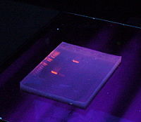
Nucleic Acid Gels Stain
Nucleic acid gels are commonly stained with fluorescent dyes or chemical stains to visualize DNA or RNA bands after gel electrophoresis.
Some popular stains include:
Ethidium Bromide (EtBr):
It's one of the most commonly used stains. Ethidium bromide intercalates between the base pairs of DNA and fluoresces orange-red under UV light. However, it's toxic and a potential mutagen.

SYBR® Safe:
It's a safer alternative to EtBr. SYBR Safe also intercalates with DNA but is less toxic and can be used in place of EtBr with similar sensitivity.
GelRed™ and GelGreen™:
These are other examples of safer alternatives to EtBr. They are also fluorescent stains and have similar sensitivities.
SYBR® Green:

It's a highly sensitive stain that binds to DNA but does not intercalate between base pairs. It's less mutagenic than EtBr but more expensive.
Methylene Blue:
This stain binds to DNA but does not fluoresce under UV light. Instead, it gives a blue color to DNA bands. It's less sensitive compared to fluorescent stains but is less toxic.
Silver Staining:
This method involves staining DNA with silver ions, resulting in the deposition of metallic silver onto the DNA bands. It provides high sensitivity and is commonly used for protein gels as well.
Coomassie Brilliant Blue:
Although primarily used for protein staining, it can also stain nucleic acids. However, it's less sensitive than other methods and usually requires post-staining with silver.
These stains have different sensitivities, ease of use, and safety profiles, so the choice depends on factors such as the specific application, budget, and safety concerns. Additionally, newer stains and techniques continue to be developed to address issues such as sensitivity, toxicity, and environmental impact.
